
our services
Breast Imaging & Intervention
At Diagnostic Imaging for Women we use the most up-to-date radiography equipment to ensure comfort, ease and accuracy for our patients.
Breast imaging provides a detailed look into the breast tissue and allows our team to best support your health.
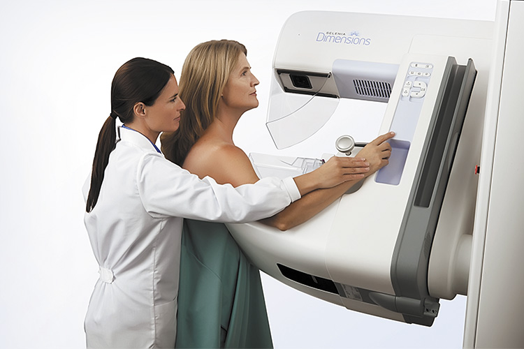
Digital & 3D Mammogram
Mammograms are vital in assessing the health of breast tissue.
Your first mammogram is considered a baseline mammogram against which all future tests will be compared to look for changes in your breast tissue.
There are two types of mammograms – screening and diagnostic.
our approach to mammograms
The standard imaging at Diagnostic Imaging for Women consists of 2D mammography in conjunction with 3D mammography. 2D mammograms capture images of the breast from the top and from the side, whilst 3D mammograms (or tomosynthesis) create a 3D image of the breast by capturing a series of images at various angles.
The 2D and 3D images of the breast are captured simultaneously, generating highly detailed images of the breast tissue.
This process involves 4 standard images of the breast captured from various angles. The mammographer will compress the breast gently using a paddle to hold the breast in place. Compression of the breast is necessary to ensure the breast tissue is uniform and will produce the most accurate imaging.
The benefits of combined 2D and 3D mammography are
- Reduced need for follow-up imaging
- Detection of more cancers than a standard 2D mammogram alone
- Improved breast cancer detection in dense, complex and glandular breast tissue
Mammograms can be performed comfortably and safety for patients with breast implants.
A light compression will be applied to the breast to hold the breast in place. There are 6 standard breast imaging views performed for women with implants. The first 2 views will show the implant in the breast tissue. The remaining 4 views are referred to as “pushbacks” and these involve gentle displacement of the implant to allow for compression of breast tissue only. This process ensures accurate and clear imaging of all areas of the breast that may be covered by the implant.
Due to breast tissue in younger women being denser, a combination of mammogram and ultrasound should be considered. At difw, we assess each patient’s clinical presentation individually to determine the most appropriate imaging technique.
Screening Mammograms
Screening mammograms are performed for women who have no symptoms or signs of breast cancer and are considered at average risk for breast cancer.
Diagnostic Mammograms
Diagnostic mammograms are performed for women who have symptoms such as a lump, pain, nipple thickening or discharge, or whose breasts have changed shape or size.
Core Biopsy
A core biopsy performed under ultrasound guidance may be requested by your doctor or upon radiologist recommendation if there is an area within your breast that can be seen on ultrasound and mammogram that is suspicious for either malignancy or a complex benign condition.
A core biopsy obtains small cores of tissue from an area of interest. This enables the pathologist to assess not only cells from the area but also the architecture or structure of the lesion, to assist in a definitive diagnosis.
A preliminary breast ultrasound is performed to review the area in question and allow appropriate procedural planning by our radiologist. The procedure is performed by our radiologist, with an imaging technician assisting. The radiologist will inject local anaesthetic to numb the skin surface and a deep tissue local anaesthetic to further numb the area within the breast around the lesion itself. A small incision is made in the skin through which the radiologist will pass the core needle. The core needle is then guided under ultrasound into the lesion and one or more cores of tissue are obtained. The specimen will be sent for pathology analysis.
Following the procedure, our imaging technician will apply pressure to the biopsy site to minimise bruising. A sterile dressing will be applied, and you will be consulted about aftercare.
Fine Needle Aspiration/Biopsy
A fine-needle biopsy may be requested by your doctor or upon radiologist recommendation if there is an area within your breast that can be seen on ultrasound and is suspicious for either malignancy or a complex benign condition.
An aspiration involves the removal of fluid, and may be requested or recommended if you have one or more breast cysts that have become enlarged or painful. The aspirated fluid may also be sent for pathology analysis, depending on the circumstances.
A preliminary breast ultrasound is performed to review the area in question and allow appropriate procedural planning by our radiologist. The procedure is performed by our radiologist, with an imaging technician assisting. The radiologist will inject local anaesthetic to numb the skin surface and ensure you are as comfortable as possible. Under ultrasound guidance, a fine-gauge needle is inserted into the area and reverse pressure is applied to aspirate fluid.
Following the procedure, our imaging technician will apply pressure to the insertion site where there may be a small blood spot. A sterile dressing will be applied, and you will be consulted about aftercare.
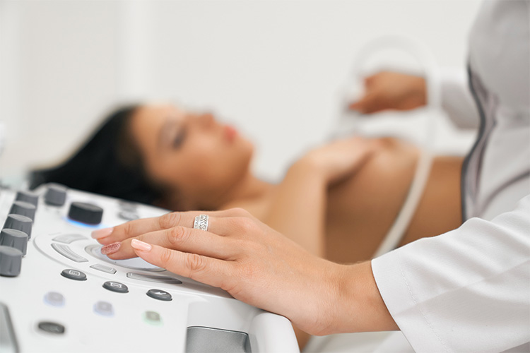
Breast Ultrasound
Due to breast tissue in younger women being denser, a combination of mammogram and ultrasound should be considered. At difw, we assess each patient’s clinical presentation individually to determine the most appropriate imaging technique.
Percutaneous Mammotomy
Percutaneous Mammotomy is a form of intervention that uses mammography technology to isolate areas in the breast such as microcalcification, that can be suspicious for ductal carcinoma in-situ (DCIS) or invasive ductal carcinoma.
A mammotomy uses a larger gauge core biopsy performed with vacuum-assistance, performed under mammographic guidance rather than ultrasound guidance. This procedure may be requested by your doctor or upon radiologist recommendation if there is an area within your breast that can be seen on mammography, but has not been detected on ultrasound, that is suspicious for malignancy or a complex benign condition.
The procedure can be performed sitting up or lying on your side, using a specially designed chair that can be adjusted to the most comfortable position.
Once the performing mammographer has you in position, compression is applied similar to a normal mammogram and preliminary images of the breast are taken. Once the performing mammographer and radiologist have targeted the area, local anaesthetic is applied to numb the skin surface. A deep tissue local anaesthetic is then also used to further numb the area within the breast around the targeted area.
A small incision is made in the skin through which the core needle will be passed using computer-generated 3D coordinates to target the area to be biopsied. Mammography images are taken throughout the procedure to document the area of interest and the specimens obtained.
Following the procedure, our imaging technician will apply pressure to the incision to minimise bruising. A sterile dressing will be applied, and you will be consulted about aftercare.