Posted on 01 April 2021
Comprehensive Breast Screenings at Diagnostic Imaging for Women - Your Questions Answered
All women aged over 40 - Although the risk of breast cancer increases significantly with age, approximately 1 in 6 breast cancers will be detected in women aged between 40 and 50. This should be followed up every 1 to 2 years. Dr Paula Sivyer will advise you of your recommended follow-up at your initial appointment.
Yes, you do. Early detection is the best protection. Our technology can detect breast cancers when they are only millimetres in size – many months before you are able to feel any lumps or experience any symptoms. Our objective is to catch breast cancers at their earliest possible stage before there are any other signs of invasive disease.
20,000 Australian women will be diagnosed with breast cancer this year alone. 9 in 10 of these women will not have a family history of the disease.
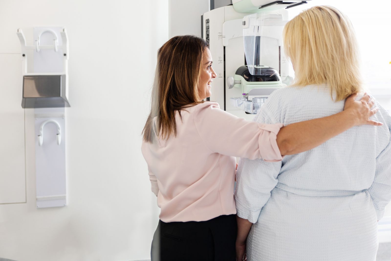
Our breast imaging encompasses mammography and ultrasound. The standard imaging protocol at Diagnostic Imaging for Women consists of 2D mammography in conjunction with 3D mammography, where a 2D image and 3D image of the breast are captured simultaneously, generating high detailed images of the breast tissue. This process involves 4 standard images of the breast captured from various angles.
The benefits of combined 2D and 3D mammography are:
- Reduced need for follow up imaging
- Detection of more cancers than a standard 2D mammogram alone
- Improved breast cancer detection in dense, complex, and glandular breast tissue
Following your mammogram, we will also conduct high-resolution breast ultrasound. These two different imaging modalities (mammogram and ultrasound) ensure a thorough and comprehensive breast health assessment. While mammography is a great diagnostic tool for breast cancer, not all breast cancers will be detected on mammogram alone.
All mammography uses x-rays, so a tiny level of radiation is required to produce the imaging. Mammography remains the gold standard for early detection of breast cancer. It is the most appropriate imaging choice for breast cancer screening in many patients. In most cases, the benefits of early breast cancer detection far outweigh the risk posed by tiny levels of radiation.
With advances in technology over the last few years, the radiation levels required to produce high resolution mammograms are at an all-time low – up to 5 times lower dose than mammograms performed 10 years ago. At difw, we are committed to using the latest technology available so that your imaging is produced at the lowest radiation dose possible.
As for high resolution ultrasound, there is no radiation dose involved.
Absolutely. We just take a couple more views. There are 6 standard breast imaging views performed for women with implants. The first 2 views will show the implant in the breast tissue. The remaining 4 views are referred to as “pushbacks” and these involve gentle displacement of the implant to allow for compression of breast tissue only. This process ensures accurate and clear imaging of all areas of the breast that may be covered by the implant.
Recent advances in technology offered by difw such as variable compression and curved paddles mean than mammography is performed with less discomfort than ever before.
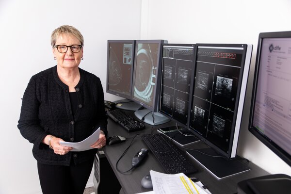
Unlike many other clinics, our consultant radiologist, Dr Paula Sivyer, will review your imaging, discuss your results and future management recommendations on completion of your appointment. This will be followed up by a comprehensive report delivered promptly to your medical team.
If further interventional procedures are required, we make the arrangements for this to occur at the earliest convenient time – often the same day. We work closely with your medical team to ensure that this process happens in the smoothest possible way, and that you are kept informed at every stage of what is going on.